A 3D Simulator of the Patellar Reflex and Demyelination Effects
You can download the simulator from the button below:
You can check the Github repo of the project from the button below:
Demo
Table of Contents
- Table of Contents
- Introduction
- Implementing The Model in Python
- Creating the 3D Visualization using Unity
- Result
This project has multiple aspects: biological/anatomical analysis, computational modeling in Python, and a 3D simulation using Unity. You can use the table of contents to navigate to the aspect that interests you most.
Introduction
This project is about building a mathematical model of the neuron and the impact of demyelination on the neuronal characteristics, more on the significance of understanding the impact of demyelination of neurons in the Demyelination of Neurons section.
To put our model in a meaningful context, we will use it to simulate the neuronal circuit of the patellar reflex. We chose the patellar reflex circuit because it’s relatively simple and doesn’t require brain processing. It can be represented by just four neurons: two motor neurons, a sensory neuron, and an inhibitory interneuron.
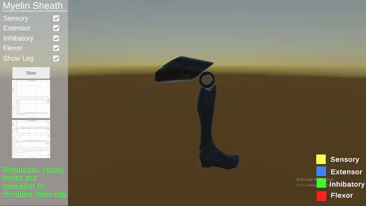
But who enjoys staring at simulation plots or a bunch of numbers? That’s why we decided to take it a step further by developing a 3D simulator, allowing the user to visualize the simulations in action on a 3D model of the leg and the neurons of the patellar reflex circuit.
Let’s start by covering the essential background you need in this introduction section.
Patellar Reflex Circuit
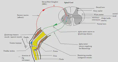
The above figure illustrates circuit diagram for the patellar reflex or knee jerk reflex which is a test used to examine the integrity of the sensory and motor pathways of a portion of the lumbar spinal cord. The tap of hammer stretches the leg muscle and leads to the initiation of action potentials in sensory neurons within the muscle that are sensitive to stretch. The action potentials propagate into the spinal cord where the axon splits into two branches. At the extensor motor neuron terminal (terminal E), the action potential propagates directly into an extensor motor neuron. This is a basic example of feedforward excitation pathway. At the interneuron terminal (terminal N), the action potential expands to an inhibitory interneuron which is interposed before the flexor motor neuron. This leads to an inhibitory postsynaptic potential which makes the action potential less likely to propagate down the flexor motor neuron so it will decrease the probability of undesired flexion in the leg muscle. This is why when a doctor taps your leg, it will extend instead of flex. This is an example of feedforward inhibition. We will be studying the effects of demyelination of these four neurons in this circuit.
The neuronal relationship of the Patellar Reflex Circuit is represented in the block diagram shown in the figure below.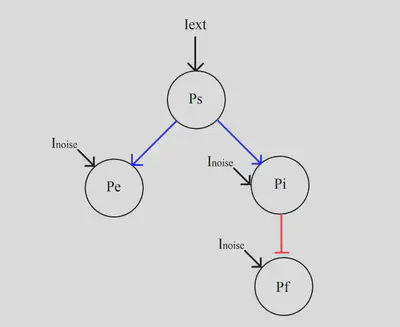
Demyelination of Neurons
The proper functioning of the patellar reflex cricuit—and indeed, the entire nervous system—relies heavily on the integrity of myelin, a fatty substance that insulates neuronal axons. Demyelination refers to the damage or loss of this myelin sheath, which can severely disrupt the transmission of electrical signals along neurons.
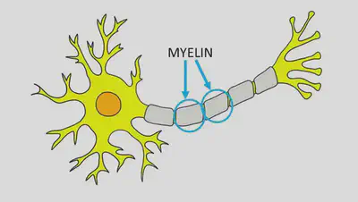
Myelin acts as an insulating layer, much like the plastic coating around an electrical wire. It allows action potentials to propagate rapidly and efficiently along the axon through a process called saltatory conduction. When myelin is damaged, this conduction slows down or fails entirely, leading to delayed or blocked signal transmission. In the context of the patellar reflex, demyelination of the sensory or motor neurons could impair the reflex response, causing either a delayed extension or a complete lack of movement in the leg.
Demyelination increases a neuron’s membrane conductance by stripping away the protective myelin sheath. This exposure raises the effective capacitance of the axon, causing slower action potential propagation and allowing more ions to leak out. As a result, the efficiency of electrical signal transmission is compromised, leading to significant functional deficits in the affected neurons.
Demyelination is a serious issue because it underlies several debilitating neurological disorders, such as multiple sclerosis (MS), Guillain-Barré syndrome, and Charcot-Marie-Tooth disease. In MS, for example, the immune system mistakenly attacks the myelin sheath in the central nervous system, leading to symptoms like muscle weakness, coordination problems, and fatigue. Over time, repeated demyelination can result in permanent damage to the axons themselves, causing irreversible neurological deficits.
The consequences of demyelination extend beyond physical symptoms. Slowed or disrupted signal transmission can affect cognitive functions, sensory processing, and even autonomic functions like heart rate and digestion. This makes demyelination not just a localized issue but a systemic one, impacting the overall functionality of the nervous system.
In this project, we explore how demyelination affects the patellar reflex circuitry by simulating the degradation of myelin in the sensory, extensor motor, interneuron, and flexor motor neurons. By understanding these effects, we can gain deeper insights into the broader implications of demyelination and its impact on neural communication.
Implementing The Model in Python
I’ve dedicated a whole blog post on modeling neurons and synaptic connections (see this post), where I included a simulation of a simple circuit of two neurons. I recommend checking it out, as I’ll be extending on the concepts and implementation discussed there.
As discussed in the Demyelination of Neurons section, demyelination increases a neuron’s membrane conductance. In our very specific case, through experimentation on the impact of capacitance on signal latency from one neuron to another, the demyelinated neuron’s membrane capacitance will increase to 7 $\mu \text{F/cm}$. This will be reflected in the value of the capacitance paramater of the neuron’s model (The Hodgkin-Huxley Model).
Building on the implementation outlined in the earlier blog post, we can extend the simulation to model the four neurons involved in the patellar reflex circuit and incorporate the effects of demyelination. Check the simulation code of patellar reflex here on Github.
Creating the 3D Visualization using Unity
Unity brings the patellar reflex to life in a fully interactive 3D simulation. It visually demonstrates how a simple tap on the patellar tendon sets off a chain reaction in the nervous system, sending signals through sensory and motor neurons and ultimately causing the leg to kick forward. More than just a basic reflex model, the project also explores how demyelination can disrupt signal transmission, providing insight into neurological conditions that affect movement. Remember to visit the Github repo to check the Unity project.
To break it down, the core of the simulation revolves around two key aspects: neural signal propagation and knee movement. The ActionPotential.cs script handles the first part, managing how signals travel along different types of neurons. It uses Transform objects to define the pathways for sensory and motor neurons, while visual effects like bloom highlights and game objects representing neural structures help bring the activity to life. An additional feature allows users to toggle myelin sheaths on and off using a checkbox, illustrating the impact of demyelination on reaction speed, visualized as a slower traveling signal that might fade out before reaching the axon terminal..
On the movement side, the KneeController.cs script ensures that the knee moves in a realistic way. It takes advantage of Unity’s HingeJoint physics component to create natural knee motion. When the neural circuit activates, the knee extends smoothly to a target angle, then gradually relaxes back to its resting position using interpolation functions. To keep things interactive, the simulation allows users to manually trigger the reflex with pressing spacebar, mimicking the hammer tap on the knee.
The project is built using standard Unity practices, keeping neural activity, movement, and user interaction neatly organized in separate scripts. With detailed 3D anatomical models enhancing the scene, the result is a compelling, educational simulation that makes an otherwise complex biological process easy to grasp.